Bioprinting
Transplants from the 3D printer
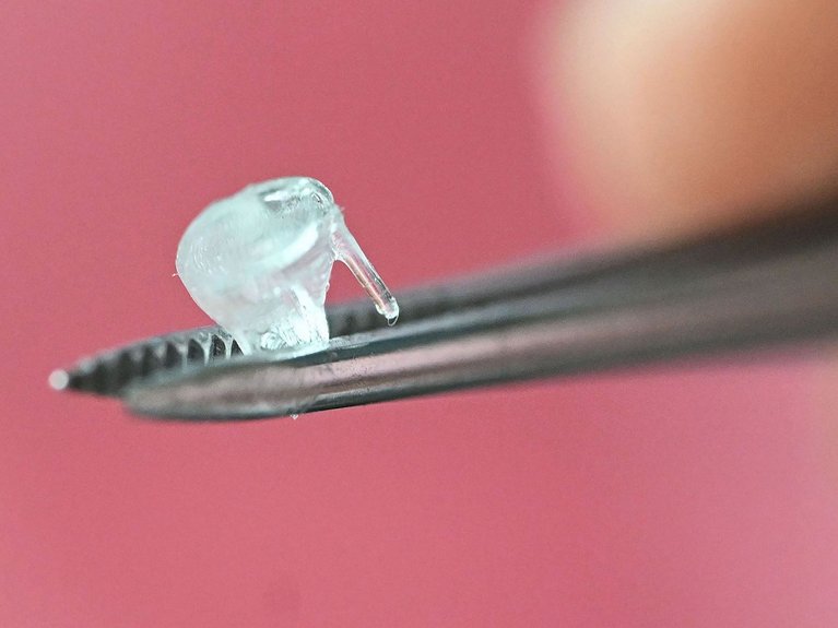
3D-printed baby heart valve. Picture: Uli Deck/dpa
At KIT in Karlsruhe, a team led by biochemist Ute Schepers is developing heart valves using a 3D printer. Bioprinting could become standard practice in transplant medicine within the next few years.
- A research team led by Ute Schepers at the Karlsruhe Institute of Technology is using a bioprinting process to create functional heart valves for babies using the patients' own cells.
- These printed heart valves grow alongside the child and are not rejected by their immune system.
- Mini-organs produced by a 3D printer can enable personalised drug testing and reduce the need for animal testing.
- Such transplants could become the clinical standard within five to ten years, and, in the long term, entire organs could be printed.
Can a new heart valve be fitted in just a few minutes? No problem! The 3D printer needed to make it is only about the size of a case of mineral water. It creates a “spare part” like this layer by layer in just 120 seconds. Researchers at the Karlsruhe Institute of Technology (KIT) designed the valve's shape and structure. It fits perfectly in the heart of a small child.
'Basically, 3D bioprinting works similarly to an inkjet printer,' says Ute Schepers, the biochemist heading the working group. The only difference is that, rather than liquid ink, the cartridges are filled with bio-ink: human body cells suspended in a collagen gel. Unlike classic collagen, this collagen, which was developed and patented by the KIT researchers in collaboration with Carl Zeiss Meditec AG and Evonik Healthcare, is not obtained from animals, but is produced with the help of bacteria. It is vegan. The scientists have also modernised the printing process, relying on Digital Light Processing (DLP), whereby a digital light projector cures the bio-ink layer by layer. This is done exactly according to a previously created 3D model.
Artificial heart valves are urgently needed. In Germany alone, thousands of babies are born with heart valve malformations every year. In some cases, their hearts cannot pump blood through the body with enough force. This puts extra strain on the organ, causing it to weaken. "In such cases, a transplant is often the only solution," says Ute Schepers.
Until now, most heart valve transplants have come from pigs. The main disadvantage is that they do not grow alongside the recipient's body. This is because the operation causes scarring and calcification, which stops the cells in the tissue from dividing in an unfamiliar environment. Children affected by this therefore have to undergo multiple operations and be fitted with new heart valves repeatedly until their heart reaches its final size.
The heart valves from KIT are intended to eliminate the need for such additional surgeries. The principle behind this is as simple as it is ingenious: stem cells obtained from the skin of the child due to receive the transplant are used to produce a valve in a 3D printer. Not only did the researchers led by Ute Schepers succeed in cultivating and multiplying these body cells, they also managed to... After the operation, they continue to divide, enabling the valve to grow with the child. Another advantage is that, as the transplant does not contain any foreign cells, it is not attacked by the child's immune system. At least, that's the theory.

Prof. Dr. Ute Schepers, Department leader Chemical Biology at Karlsruher Institut für Technologie. Bild KIT
Across the globe, an increasing number of research groups, pharmaceutical companies and start-ups are using 3D bioprinters to create tissue and organs from human cells. Stem cells can be obtained from a small skin sample or umbilical cord blood and converted into other cell types in the laboratory. These cells can then be used to produce the desired tissue.
However, there are still some hurdles to overcome. For example, KIT researchers led by Ute Schepers are now collaborating with clinicians to test the durability of heart valves produced by a 3D bioprinter in the human body's chemical and physical conditions using surgical training devices. Ute Schepers is confident. "Replacement parts from the 3D bioprinter could become standard in human medicine," she says. In the case of heart valves, this could happen within the next five to ten years. In the medium term, entire hearts could also be produced using the printer. Schepers has another application for her research in mind: mini-organs produced by the 3D printer could be used to test medicine ingredients.
"This method can already be used today to avoid or at least reduce animal testing," says the biochemist. “And I see enormous potential for the future.” An additional advantage of such tissue-based tests is that medicine is increasingly recognising that active ingredients and drugs do not affect everyone in the same way. Individual differences in genetic makeup appear to be a particularly decisive factor in this regard. However, mini-organ systems printed from bio-ink containing a person's own body cells can quickly and specifically test which drugs are particularly effective for that person. This could be used to treat kidney disease, cardiac arrhythmia, or cancer, for example.


Readers comments