Robots in the blood
Fighting cancer at the root

Among other things, remote-controlled microrobots could deliver drugs to specific tumor sites. Image: Gorodenkoff/Shutterstock
“A major problem in cancer medicine today is the very low efficiency,” says Tian Qiu, who heads the Smart Technologies for Tumor Therapy working division at the DKFZ in Dresden. “With conventional chemotherapy, only one percent of the drug actually reaches the cancer site. The remaining 99 percent causes side effects somewhere else in the body." One solution would be to target the drugs precisely to the tumor. And that is exactly what he and his team are trying to do. “We are developing tiny robots for various medical applications,” he explains. “From the millimeter to the micrometer to the nanometer range.” These mini robots can be controlled wirelessly, for example using a magnetic field, or with ultrasound. “We can control where they move and recognize where they are. And we can trigger functions - such as releasing medication or taking pictures.”
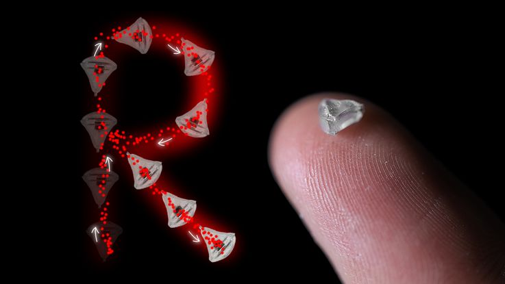
Tiny little things for the journey through the body: On the right a miniature robot with built-in SMOL tracker, the built-in magnet is only 1 mm in size. On th left an R-shaped movement path of a SMOL-controlled miniature robot. Picture: Qiu/DKFZ
Each of these three size scales is predestined for a different medical purpose. The smallest are the nanobots. Measuring just a few hundred nanometers, they are similar in size to many viruses. “They can be used, for example, to deliver drugs to a specific cancer site,” says the researcher. “The active ingredients are coupled to the surface of the tiny particles. These can move actively in the body and release the drug at the right place.” The microbots are a good thousand times larger. At a few hundred micrometers, they are as thick as a human hair and are therefore visible to the naked eye. And they can be equipped with tools. “We have developed VIBEBOT in this class. That stands for Vibrational Microrobots in Viscoelastic Biological Tissues. It’s a micro-robot that can work in the brain, for example.” The largest robots in Tian Qiu’s repertoire measure just a few millimeters. They also carry tools. A cannula, for example, or a camera, or a surgical instrument. “It is difficult for today’s medical instruments, such as endoscopes, to reach certain places in the body. For example, to perform an operation on a tumor hard-to-reach,” he explains. “Our submillimeter robots can help transport an endoscope; to get images of the tumor. Or even to perform an operation. And all minimally invasive.”
Inspired by sperm
In 2019, the DKFZ established a branch office in Dresden to drive such research forward. And in June 2023, Tian Qiu and his group moved from Stuttgart to the Saxon state capital to set up his new department. The tiny robots have been following the researcher since he studied at Tsinghua University in Beijing. There he studied microfluidics - the way in which fluids behave in tiny channels. He mainly investigated this using sperm cells. “On the one hand, they can swim in bodily fluids,” he says. “But what is even more astonishing is that they can perceive the gradient of the chemicals secreted by the egg cells.” They therefore pick up the chemical trail of the egg cells and move towards them. “As an engineer, I thought to myself: it would be super cool to develop an artificial robot that can swim in the body and has certain intelligence,” says Tian Qiu. Because there, he thought, the machine could follow a concentration gradient of certain chemicals and perform useful biomedical tasks at its destination. “That inspired me. And that's why I came to Europe, studied bioengineering in Lausanne and have been researching small robots ever since.”

Prof. Dr. Tian Qiu, Head of the department Smart Technologies for Tumor Therapy at the German Cancer Research Center (DKFZ) in Dresden. Picture: M.Stark/DKFZ
One major challenge lies in localizing the tiny robots in deep tissue. In some experiments, the researchers circumvented the problem by opting for the vitreous body of the eye. “It is transparent, so we can see our robots under the light microscope,” explains Tian Qiu. “But the other tissues in the body is not transparent. And it's very difficult to get a real-time picture of what's going on there.” Of course, there are advanced medical imaging techniques such as magnetic resonance imaging (MRI) or computed tomography (CT). But these are not ideal for small-scale robots. This is because the magnetic fields interfere with MRI. And CT involves radiation, which researchers want to avoid. In addition, these methods cannot be used to scan in real time. But this is necessary to control the robot. “For this reason, we have developed our own technology for tracking,” says the researcher.
The miniaturized tracker was built by Tian Qiu’s doctoral student Felix Fisher, published recently in the journal npj Robotics. It is a small permanent magnet that is made to vibrate. This generates a magnetic signal that can be detected by a sensor array. “The magnet is smaller than a millimeter and sits at the tip of a cantilever,” explains Tian Qiu. “It has an oscillation frequency of 135 Hertz and generates a complicated magnetic field.” The researchers use a computer algorithm to fit the magnetic field so that they can localize the tracker. “So we know where this little tracker is, and in all six degrees of freedom.” This means that they can not only detect its movement in three-dimensional space, but also know the angle of rotation around each of these axes. They have also chosen the frequency carefully. “Our power grid here in Germany vibrates at 50 hertz. This generates magnetic noise,” he explains. “It’s similar with the earth’s static magnetic field. But none of that matters if we work with 135 Hertz.” The system can report where the robot is in the body almost in real time. It delivers around five images per second. Compared to modern video technology, this is not much. But it is sufficient for minimally invasive surgical instruments. This is because they move relatively slowly in the body. “We can simply attach our small tracker to the tip of an endoscope,” says Tian Qiu. “Then we can track where the endoscope is moving in real time, and without any radiation.”
Movement in difficult terrain
However, before the tiny machines become part of the standard medical repertoire, the researchers still have a few challenges to overcome. The biggest one for Tian Qiu is penetrating the biological barrier. He starts a video of a tiny robot moving elegantly through a liquid. “We have developed the microbots over the last few years,” he says, emphasizing that it works very well in Newtonian fluids. These are liquids such as water, for example, whose viscosity is independent of the forces at work. However, this does not apply to body fluids such as blood. “That’s why we have a problem when we inject the robots into real biological tissue,” continues Tian Qiu. “They get stuck in such a biological environment.” He starts a second video. The tiny robot is now just spinning in circles. Progress is out of the question. “A solid tumor is soft tissue. And the mucus layer over almost every organ is not only water. They are viscoelastic biological fluids,” he says. “Basically, wherever we want our robots to do something useful, we come up against this barrier.” One of Tian Qiu's most important tasks is to make the robots able to move in real tissue.


Readers comments